The Cervical Axis Protocol: Tech-Neck, Muscle Guarding & the Postural Paradox
For modern corporate tech professionals, prolonged screen engagement forces severe mechanical imbalances down the cervical spine. Explore how structural curve variants, neurological mapping, and involuntary cellular defense mechanisms keep your body trapped in chronic distress.
A 34-year-old software engineer from HITEC City walked into our clinic with a 6-month-old MRI report, three failed physiotherapy courses behind him, and one persistent, frustrated question: "Why does relaxing on my soft sofa make my neck pain instantly worse, while sitting at my office desk feels better?"
For the modern technology professional, prolonged screen engagement is not merely a career reality; it is a profound physical burden on the cervical spine. In clinical practice, we continuously consult with software engineers, data analysts, and corporate executives who present with chronic, unyielding neck pain. Many of these individuals arrive with advanced MRI reports detailing multi-level disc bulges, osteophyte formation, and structural shifts. Yet, they are frequently left confused as to why their pain behaves so unpredictably flaring up during a relaxing evening on a sofa, or causing a deep, traveling ache down the arm that refuses to resolve.
To break this cycle of structural strain, we must look past superficial symptoms and decode the complex interplay of cervical alignment, neural pain pathways, and localized muscular defense mechanisms. Just like our previous findings on Ayurvedic management for back pain in Hyderabad, long-term relief requires shifting our focus away from the localized symptom and toward the entire integrated physical chain.
Part 1: The Structural Matrix: Normal vs. Lost Cervical Lordosis
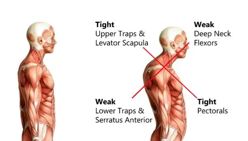
The human neck is engineered with a natural, elegant forward C-shaped curve known as cervical lordosis. This curvature functions as a built-in suspension system, distributing the gravitational weight of the head evenly across the cervical vertebrae and cushioning the intervertebral discs from daily mechanical shocks. When a professional spends hours slouched over a laptop, peering at dual monitors, or tracking code on a mobile device, this alignment undergoes a significant mechanical shift.
1. Chronic Mechanical Loss of Lordosis
Continuous forward-head posture forces the cervical spine to flatten out, leading to a "straightened" neck or, in advanced cases, a reversed kyphotic curve. When you lose this natural curve, the mechanical load on the lower cervical segments (C5-C6 and C6-C7) increases exponentially. For every single inch your head drifts forward, an extra 4.5 to 5 kilograms of baseline pressure is forced directly onto the delicate lower discs and facet joints.
2. The Techie Degeneration Sequence
Under this relentless, uneven weight distribution, the body initiates a structural stabilization response. The intervertebral discs begin to lose water content and deflate (disc desiccation). To prevent the unstable joint axis from shifting further, the borders of the vertebrae begin to produce defensive bone spurs known as osteophytes. This degenerative sequence can manifest in two distinct clinical profiles:
- Degeneration with Loss of Lordosis: The spine is structurally flattened, the discs are compressed, and the joints are rigidly locked in a forward-leaning posture.
- Degeneration without Loss of Lordosis: The natural C-curve is radiologically intact on an MRI, yet specific localized joints are suffering from severe, micro-mechanical wear and tear due to repetitive ergonomic stress.
Part 2: Neurological Mapping: Centralized vs. Decentralized Pain
When evaluating cervical disc bulges and osteophyte tracking, understanding the directional behavior of your pain is crucial for determining the stage of structural injury.
Centralized Cervical Pain
This pain pattern remains localized strictly within the midline of the neck or the immediate base of the skull. Centralized pain is typically discogenic or joint-driven. It indicates that while the outer walls of the cervical discs are under immense mechanical duress or suffering from micro-tears, the internal gel core has not yet encroached upon the primary spinal nerve roots.
Decentralized Pain & Radiculopathy (Peripheralization)
When a cervical disc bulge extends outward or an osteophyte directly reduces the diameter of the neural pathways (foraminal narrowing), it begins to physically pinch or chemically irritate the exiting spinal nerve root. This triggers a decentralized tracking pattern. The pain breaks out of the neck and travels along the nerve gateway, radiating deeply into the shoulder blade, down the deltoid, or down the entire arm into the fingers. This neurological presentation is known as cervical radiculopathy. Depending on the exact level of nerve compression, it presents with two distinct clinical markers:
- Bilateral or Unilateral Numbness: A persistent loss of sensation, "pins and needles," or a cold feeling traveling down specific neurological lines.
- Progressive Motor Weakness: A noticeable drop in physical capability, such as struggling to lift the arm away from the body, a weakened grip, or dropping everyday objects.
Part 3: The Postural Paradox: The Sofa vs. Upright Chair Enigma
One of the most frustrating puzzles for chronic neck pain sufferers is why certain resting positions trigger immediate agony while others provide unexpected comfort. This is not random; it is a direct reflection of how different postures alter the internal spacing of your cervical canal.
| Posture Type | Mechanical Impact on Cervical Spine | Clinical Result |
|---|---|---|
| Deep Sofa (Back Reclined) | Increases thoracic slouching; forces upper neck into extreme hyperextension. | Pinches posterior disc bulges & exiting nerve roots; triggers immediate pain. |
| Rigid Upright (Stark Straight) | Compresses facet joints directly; forces strict vertical loading. | Overloads arthritic joints; triggers severe baseline stiffness and aching. |
| Upright Relaxed (With Support) | Restores natural spinal curves; maintains centered head balance. | Relieves vertical pressure; allows the neuro-muscular layers to relax cleanly. |
Case A: The Sofa Trigger vs. Upright Relief
When you sink back into a soft, deep sofa or a heavily inclined recliner, your thoracic spine is forced into a rounded, slouched position. To keep your eyes looking forward at a screen, your lower neck is forced into flexion while your upper cervical spine is thrust into extreme hyperextension. If you have an existing posterior disc bulge or osteophyte network, this specific positioning immediately pinches the adjacent nerve root spaces, triggering a sharp wave of radiating pain or numbness within minutes. Conversely, sitting upright takes away that extreme hyperextension, opening the neural pathways and providing relief.
Case B: The Upright Trigger vs. Sofa Comfort
For a completely different structural profile, sitting rigidly upright in a standard office chair is the primary pain trigger, while leaning back into a soft sofa brings total ease. This occurs when the patient’s pain is driven by facet joint arthropathy or severe vertical compression rather than a dynamic disc bulge. Sitting starkly straight forces a direct vertical load onto compressed, worn-out joints. Leaning back into a supportive, reclined angle immediately unloads this gravitational pressure, allowing the compressed joint surfaces to separate and providing instant relief. Similar core axis failures can be reviewed in our comprehensive guide on spine surgery vs conservative care models.
Part 4: The Muscular Armor: Decoding Severe Muscle Guarding
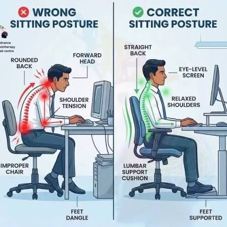
When the cervical joints and nerve pathways are under chronic mechanical stress, the central nervous system refuses to remain passive. To protect the vital spinal cord from further micro-injury, the brain commands the surrounding muscular layers to contract into a rigid, involuntary biological splint. This state of perpetual contraction is known as severe muscle guarding.
While this defense mechanism is meant to protect the spine, it quickly turns into a primary source of chronic pain and stiffness. The locked muscles completely stall localized micro-circulation, trapping metabolic waste products and starving the tissues of fresh oxygen. Depending on your specific postural habits and structural variants, this guarding reflex locks down distinct muscle groups:
- The Trapezius & Levator Scapulae: These muscles function as the heavy vertical suspension cables for your shoulder blades and neck. When lordosis is lost, they are forced to work double-time to hold the weight of a forward-drifting head. This leads to dense, burning knots, severe upper shoulder tightness, and a constant feeling of exhaustion across the top of the shoulders.
- The Rhomboids: Located directly between your shoulder blades, the rhomboids continuously fight against the forward-rolling shoulder pattern common among desk workers. Under chronic strain, they develop a deep, nagging, localized ache that patients frequently try to rub out against a wall or chair back.
- The Sternocleidomastoid (SCM) & Splenius Capitis: The SCM muscles run along the front and sides of the neck, while the splenius capitis controls the back of the skull. When these muscles lock up to guard the spine, they restrict your ability to turn your head from side to side, causing deep stiffness and triggering tension headaches that radiate behind the eyes or across the temples.
Part 5: The Commuter’s Matrix: Cervicogenic Vertigo and Screen-Induced Motion Sickness
A significant segment of technology professionals presents with a highly distressing symptom footprint that surfaces exclusively during travel: the instant they look down at a mobile device or laptop inside a moving vehicle, they experience an immediate loss of balance, spatial disorientation, dizziness, and intense, wave-like nausea. While these symptoms perfectly mimic inner-ear disorders like Benign Paroxysmal Positional Vertigo (BPPV), their true origin lies in a profound breakdown of the communication lines between the upper cervical spine, the eyes, and the brain.
1. The Tri-Unite Sensory System
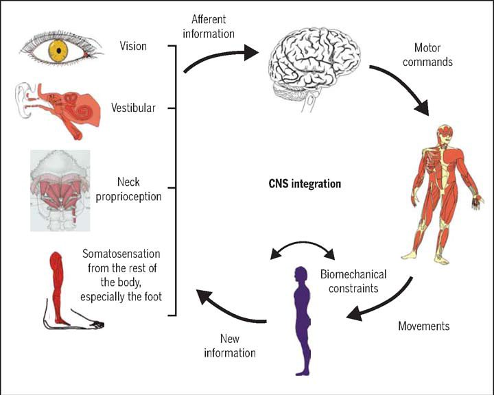
To maintain perfect balance and orientation, your brain continuously relies on a real-time stream of data from three distinct sources: the vestibular system (the fluid-filled canals of your inner ear), the visual system (your eyes tracking your environment), and the proprioceptive system (the deep suboccipital muscles and upper cervical joints C1, C2, and C3, which possess the highest density of spatial position-sensing receptors in the human body).
2. The Mechanics of a Sensory Conflict
When an individual with chronic tech-neck degeneration and severe muscle guarding slumps over a screen in a moving car, a catastrophic data mismatch occurs. Your inner ear senses the physical acceleration, bumps, and turns of the vehicle. Your eyes, locked onto a static phone screen, report to the brain that your body is completely stationary. Simultaneously, the chronically tight, guarded muscles of the upper neck specifically the SCM and Splenius Capitis are under extreme static strain, sending distorted, locked proprioceptive signals to the central nervous system.
The brain suddenly receives three completely conflicting sets of data. It cannot determine where the head is relative to the moving environment. This sensory chaos instantly compromises the neural gateways, forcing the autonomic nervous system into an emergency distress response. The direct result is a sudden onset of cervicogenic dizziness, spatial disorientation, balance failure, and systemic nausea.
The Timeline Reality: Why Complex Cervical Cases Outlast a 14-Day Window
One of the most common misconceptions in spinal rehabilitation is the belief that chronic, multi-level cervical disc issues and sensory disorders can be permanently resolved within a standard 14-day treatment window. While a two-week intensive course of specialized clinical therapy can achieve remarkable milestones, expecting an absolute structural reset in this timeframe is anatomically unrealistic for complex cases.
When a patient presents with a clean, uncomplicated muscular strain, a 14-day protocol is often more than enough to restore complete mobility. However, when a spine is navigating a multi-layered failure such as a multi-year history of structural degeneration, loss of cervical lordosis, open annular tears, or chronic sensory mismatch loops causing vertigo the clinical clock operates differently. For these intricate cases, recovery must be viewed as a multi-stage journey, not a quick two-week fix:
- Phase 1 (Days 1 to 14) Neural Desensitization & Gate Control: This initial window is highly effective for overriding the neural guarding reflex, flushing out localized fluid stagnation, and clearing static nerve blocks. It re-opens the space around the nerve roots, which often brings a significant drop in active pain intensity, dizziness, and immediatemuscular tightness.
- Phase 2 (Weeks 3 to 12+) Tissue Regeneration & Mechanical Unloading: Clearing active nerve pain does not mean the underlying structural breach has magically healed. Compressed disc walls, dehydrated joint spaces, and strained suboccipital muscle spindles require months of steady micro-circulation to rebuild and stabilize. If a patient has an under-muscled frame or uncorrected metabolic structural depletions, the tissue lacks the baseline resilience to hold a correction without extended time and nutritional support.
- Phase 3 (Long-Term) Postural Discipline & Functional Resilience: Once the initial 14-day clinical window closes, the cervical axis is highly vulnerable. If a patient immediately discontinues care, skips their prescriptive neck isometric movements, and returns to a rigorous corporate routine sitting slouched over a screen for 12 hours a day or reading on phones during bumpy commutes the unstable joints will immediately buckle under gravity. The nervous system will instantly reactivate its painful protective muscle splint, leading to an immediate relapse.
To put it plainly: We cannot reverse years of progressive mechanical degradation, systemic depletion, and unmitigated screen stress in just 14 days. Our clinical protocols are designed to aggressively kickstart your recovery and clear your immediate neural pain blocks within that initial window. However, achieving permanent, unshakeable stability requires a committed, extended timeline of lifestyle corrections, targeted functional rehabilitation, and steady postural discipline.
Part 6: The Vedansha Neuro-Mechanical Framework
Lasting structural recovery requires a comprehensive approach that targets the nervous system, the deep guarding muscles, and the weight-bearing joint axis at the same time. You can learn more about how this mechanism works in our breakdown of the core benefits of Marma therapy.
- Resetting the Neural Gate: We begin by applying precise, gentle manual pressure onto key neuro-receptive vital gateways across the cervical, scapular, and cranial regions, such as the Krichatika Marma. These precise stimulations target the Gate Control Theory of Pain at a cellular level, sending immediate, high-velocity sensory signals up the thick nerve fibers to the spinal cord. This closes the neural gateway to pain, effectively silencing hyperactive nociceptors and commanding the brain to deactivate its painful protective muscle splint.
- Clearing the Guarding Layer via Muscle Manipulation & Effleurage: Once the brain drops its defensive guard reflex, we introduce deep, targeted muscle manipulation and specialized manual effleurage sequences. These fluid, directional strokes are specifically designed to break open dense muscle knots, release chronic tissue tension, and stretch shortened muscle fibers across the trapezius, levator scapulae, rhomboids, and SCM groups without triggering a defensive reflex.
- Activating Tissue Decongestion via Lymphatic Drainage: Chronic muscle guarding traps metabolic inflammatory fluids around the cervical nerve roots and suboccipital junctions, sustaining a state of chemical irritation and dizziness. By integrating precise manual lymphatic drainage techniques, we physically guide this stagnant fluid away from the compressed spinal gateways and into the body's natural circulatory clearance pathways. This non-invasive clearance immediately reduces local swelling, improves tissue elasticity, and provides the healthy, oxygen-rich environment required for compressed disc walls to naturally recover their resilience.
- Building Long-Term Postural Endurance: True stabilization requires active patient partnership. Once manual therapies have successfully cleared the nerve compression and restored joint tracking, we guide you through precise neck isometric exercises designed to build deep cervical muscle endurance, protecting your structural axis during everyday corporate activities.

Tired of Your Neck Stiffness or Commuter Dizziness Returning Post-Treatment?
Stop settling for short-term symptomatic relief. Schedule a comprehensive, doctor-led cervical axis assessment and clinical Marma evaluation in Hyderabad today, and let's fix the true mechanical root cause of your pain.
Book Your AssessmentDisclaimer: This article is for informational purposes only and does not constitute formal medical advice. Please consult a qualified healthcare professional for precise clinical evaluation and treatment mapping.